 Techcomp headquarters
  Techcomp regional offices
  Manufacturing, design and R&D facilities

product
- Atomic Force Microscope
- FE-SEM
- Hi-SEM
- Optical Interferometry System
- Sample Preparation
- Tabletop Microscope
- Transmission Electronic Microscope
- X-Ray Spectroscopy
- Fluorescence Spectrophotometer
- Amino Acid Analyzer
- Atomic Absorption Spectrophotometer
- Autotitrator
- Mercury Analyzer
- UV-VIS Spectrophotometer
- High Performance Liquid Chromatography
- Thermal Analyzer
- Automatic Moisture and Ash Content Analyzer
- Fluorescence Spectrophotometer
- Fluorescence Steady State & Life Time / Flash Photolysis
- Mercury Analyzer
- Raman Spectroscopy
- UV-VIS Spectrophotometer
- Mass Spectrometer
- FTIR Spectrometer
- Chromatography Data System
- Gas Chromatography
- Gas Chromatography-Mass Spectrometer (GC-MS)
- Liquid Chromatography
- Sampler and sample pretreatment system
- Column and Consumable
- Amino Acid Analyzer
- Anaerobic & Hypoxic Chambers
- Autoclave & Sterilization
- Biological Safety Cabinets & Clean Benches
- Blood bank & Phamacy Freezer
- Cryopreservation Systems
- DNA & RNA Purification
- Environmental Chambers
- Freezer Dryer
- High Capacity Centrifuge
- High Speed Centrifuge
- Lab Water Purification
- Lab Oven & Incubator
- Life Science
- Mixer
- Pipette
- Tabletop Centrifuge
- Ultracentrifuge
- ULT Freezer
- Temperature Forcing System
- Precisa Analytical Balances
- Precisa Micro Balances
- Precisa Precision Balances 0.001g
- Precisa 520 PB/PT Analytical and Precisa balances
- Preicsa 320 XB Series Balances
- Precisa 165 BJ Series Balances
- Precisa 410 SRS/SRC Series Scale
- Precisa 365 EM/330 XM Moisture Analyzers
- Precisa 490 Series Industrial Scales
- Precisa 321 LX/LS-STB Series Stirrer Balances
- Precisa 321 LG Series Balances
- Standard Balances
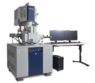
Hitachi Ultrahigh-Resolution Scanning Electron Microscope SU8600
The SU8600 brings in a new era of
ultrahigh-resolution cold-field emission scanning electron microscopes to the
long-standing Hitachi EM lineup. This revolutionary CFE-SEM platform
incorporates multifaceted imaging, automation, increased system stability,
efficient workflows for users of all experience levels, and more.
- Hong Kong
Hitachi Ultrahigh-Resolution Scanning
Electron Microscope SU8600

UltraHigh-Resolution
Hitachi’s high-brightness cold field emission source provides ultrahigh-resolution images even at Ultra-low voltages.


RHO-type Zeolite particle at low-kV. In order to reveal fine steps structure on surface, the image was acquired at 0.8 kV of landing voltage. This allows the very fine structure of surface steps to be clearly visible (image on right).
A Smart Detection System for Low Voltage BSE Imaging
Cross section image of 3D NAND;
Oxide layer and Nitride layer of capacitor are
easily distinguishable in the image due to BSE detection capability.

Cross Section of 3D NAND (Acceleration Voltage: 1.5 kV)
Fast BSE Imaging : New Out-Column Crystal Type BSED (OCD)
By using new Out-Column Crystal Type BSED (OCD)*,
image acquisition time was less than ONE SECOND, yet lower layer interconnect
and Fin FET structure of SRAM are clearly visible.

Lower Layer Interconnect of 5 nm process SRAM (Acceleration Voltage: 30 kV, Acquisition time <1 second)
Enhanced User Experience with Advanced Automation
The “EM Flow Creator“ software option allows users to configure repeatable SEM operation sequences.
Various SEM functions can be assembled in the EM Flow Creator’s window by a drag-and-drop method and then saved as a recipe for later use.
Once a recipe is configured, automated data collection under the set conditions can be performed with high accuracy and repeatability.

Flexible
Interface
Dual monitor configuration supports a flexible and highly efficient workspace. Display and save 6 signals simultaneously in order to acquire more information in less time

More information about Hitachi Scanning Electron
Microscope SU8600, please visit Hitachi High Tech official site at link:
https://www.hitachi-hightech.com/global/en/products/microscopes/sem-tem-stem/fe-sem/su8600.html
Application:
Semi-conductor, Nano Material, Advance Material

 2606, 26/F., Tower 1, Ever Gain Plaza, 88 Container Port Road, Kwai Chung, N.T., Hong Kong
 +852-27519488 / WhatsApp/WeChat HK: +852-8491 7250
 techcomp@techcomp.com.hk
 Sweep The Concern Us